
Contrast Radiology
What does it consist on?
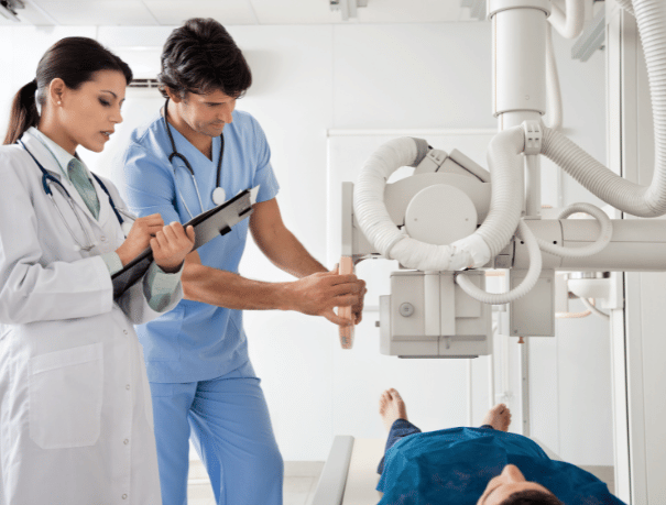
This technique is useful for evaluating the state and functioning of the different structures inside the human body. For this, a fluorescence screen with X-rays and contrast media, such as iodine and barium, are used. These are administered intravenously, or they are ingested or introduced as an enema, depending on each case, which makes it easier to visualize the organs and structures.
Cases in which it is recommended
To whom?
The doctor may ask you to perform a contrast X-ray to rule out and diagnose possible pathologies that affect the following organs and structures:
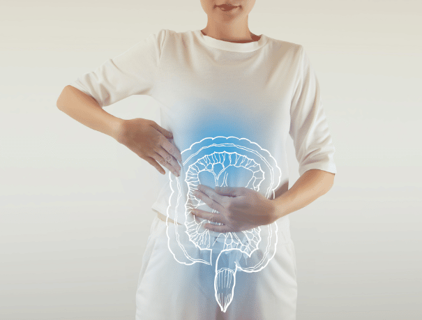
- Esophagus, stomach, small intestine and large intestine.
- Fallopian tubes (if they are blocked or inflamed) and the uterus (size, shape, structure).
- Ducts of the parotid and submandibular salivary glands.
- Bladder (structure and shape)
- Urethra, in order to detect possible narrowing of the lower urinary tract)
- Kidneys, bladder, and ureters (the tubes that carry urine from the kidneys to the bladder).
- Orifice of the fistula, in order to demonstrate the existence of the fistula and its morphology.
Instructions
How should you prepare?
- Intestinal transit: Go fasting for 6 hours.
See preparation Hysterosalpingography
- Sialography: Bring a lemon and a belt.
See preparation Colonic Transit

Medical professionals
The specialists who will assist you at CreuBlanca
A team of professionals to take care of you.


CreuBlanca centers
Find your nearest CreuBlanca center

Diagnosis Médica
Related articles
CreuBlanca's blog
You will find advice from our professionals on how to improve your health and information on the latest technologies applied in the medical health sector.
 Specialties
Cystitis and thrush in summer: how to tell them apart
 Specialties
Elevated PSA: the imaging that changes everything
 Specialties