
Osteoarticular CT
What does it consist of?
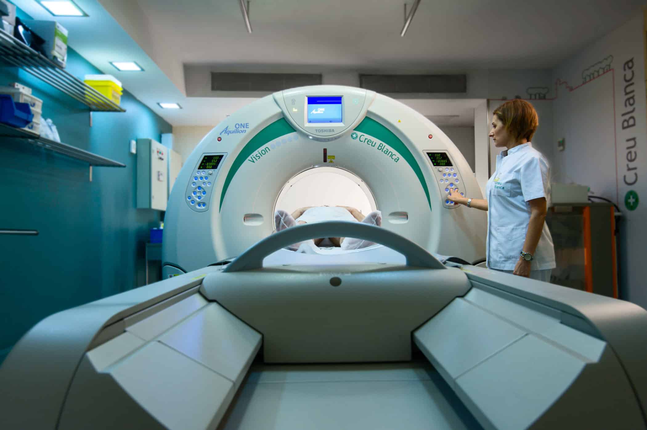
Once you have been placed on the CT table, it will begin to rotate around your body to obtain cross-sectional projections of bones, blood vessels and soft tissues, which are subsequently grouped using Artificial Intelligence. Instead of obtaining a single image, as occurs with conventional radiography, CT obtains multiple images with higher quality and sharpness, which increases diagnostic precision.
TYPES OF OSTEOARTICULAR CT
What explorations do we perform?
- Arm CT. Study of the area that includes the upper and lower part of the arm.
- Shoulder CT. Explore the shoulder area and the joints that surround it.
- Clavicle CT. Examine possible cracks or breaks, among other injuries, in the clavicle bone.
- Hand CT. Explore possible injuries to ligaments, tendons, and small joints in the hand.
- Wrist CT. Study of the wrist bone and the joints that surround it.
- Finger CT. It allows to diagnose frequent capsulitis due to trauma, tendon ruptures and osteoarthritis.
- Maxillary CT. Three-dimensional study of the maxillary bones, the pieces of bone or cartilage, in which the teeth are embedded.
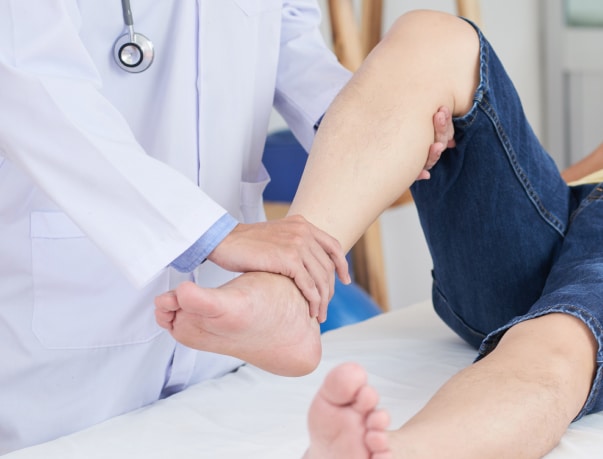
- Knee CT. Explore possible knee ligament and tendon injuries.
- Spine CT. Explore possible injuries in the cervical, lumbar and dorsal area.
Instructions
How should you prepare?
- Contrast administration: Most CT tests require the administration of contrast, either orally or through a vein. This allows the internal structures of the body to be visualized more clearly.
- Hydration: It is recommended that you come to the test well hydrated, having drunk at least one liter of water during the ten hours prior to the study. This makes it easier to remove contrast from the body.
- Metal objects: The technician will give you the necessary instructions, provide you with a gown, and ask you to remove any metal objects (jewelry, watches, piercings, hairpins, cell phones, dentures, and hearing aids).
- Cardiac devices: It is important that you inform the technician about any devices that you have implanted in your body (pacemakers, electrodes, and clips from previous surgeries). It is important that you inform the technician about any devices that you have implanted in your body (pacemakers, electrodes and clips from previous surgeries).

Medical professionals
The specialists who will assist you at CreuBlanca
Un equip de professionals per cuidar de tu.


CreuBlanca centers
Find your nearest CreuBlanca center

Barcelona
Clinic CreuBlanca

Mataró
CreuBlanca Maresme Hospital
Related articles
CreuBlanca's blog
You will find advice from our professionals on how to improve your health and information on the latest technologies applied in the medical health sector.
 Specialties
02 Jul 2026
3 Min
Cystitis and thrush in summer: how to tell them apart
Heat, humidity and long hours in a wet swimsuit make two very common conditions more frequent in summer: cystitis and vaginal thrush (candidiasis). Both affect the intimate area, both get worse with heat, and that is why many women confuse them.
 Specialties
22 Jun 2026
3 Min
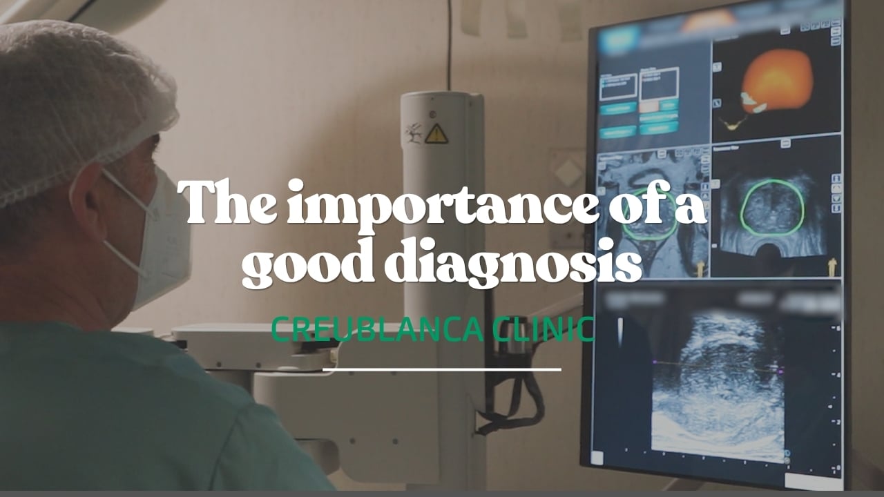
Elevated PSA: the imaging that changes everything
Elevated PSA.
No pain, no symptoms, and no obvious signs that something might be wrong.
 Specialties
28 May 2026
3 Min
Stiffness in the hands and fingers: causes, treatment and rehabilitation
Hand and finger stiffness may be caused by osteoarthritis, injuries, scars or conditions. Learn when to seek medical advice and how rehabilitation can help.