
The test
What does a functional magnetic resonance imaging (fMRI) scan involve?
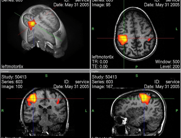
The fMRI study is based on obtaining brain activation maps by performing specific tasks during the examination. Unlike other MRI scans, not only anatomical images are acquired, but also functional information that allows precise identification of which areas of the brain are involved in specific functions.
At our center, we perform targeted studies that include:
- Motor mapping: activation of the areas responsible for movement.
- Language mapping: localization of the areas involved in speech production and verbal comprehension.
- Sensory mapping: identification of the regions related to body sensation.
These studies make it possible to establish the relationship between a lesion (tumor, vascular malformation, etc.) and the functional areas of the brain.
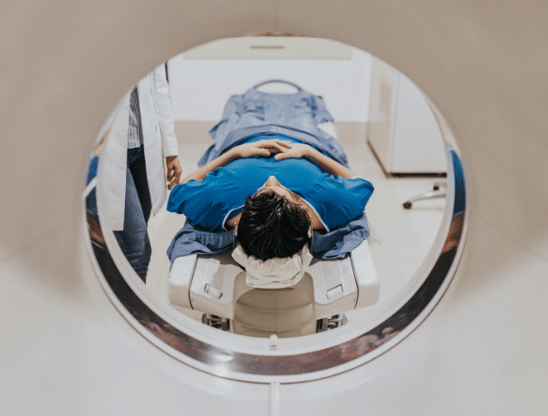
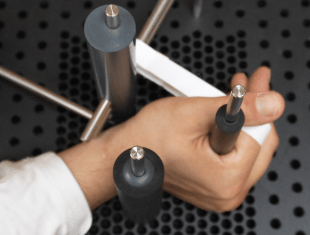
During the examination, the patient lies on their back inside the MRI scanner. They will be asked to perform a series of simple tasks (such as moving a hand, naming objects, or responding to stimuli) at specific moments following instructions. The examination usually lasts between 40 and 60 minutes, with rest intervals. Once completed, the patient can resume their normal daily activities.
CASES IN WHICH IT IS RECOMMENDED
Who is it for?
Functional magnetic resonance imaging is indicated in multiple situations, especially when it is necessary to determine the exact location of brain functions:
- Pre-surgical planning of brain tumors and other intracranial lesions.
- Assessment prior to embolization of cerebral vascular malformations.
- Study of the relationship between lesions and eloquent areas (motor, language, or sensory regions).
- Functional evaluation in patients at risk of neurological deficits associated with a structural lesion.
- Support in therapeutic decision-making and personalized treatment planning.
INSTRUCTIONS
How should you prepare?
- Preliminary interview 2–3 days before the examination: the physician responsible will meet with the patient beforehand to explain the procedure and the tasks they will be asked to perform.
- Preparation: in most cases, no special preparation or contrast administration is required.
- During the examination: it is essential to remain as still as possible and follow the team’s instructions (movements, language tasks, sensory stimuli).
- Metal objects: you must remove any metal objects (jewelry, watches, mobile phones, removable prostheses, etc.).
- Implanted devices: always inform the team if you have a pacemaker or any other medical device.
- Duration: approximately 20 to 40 minutes, depending on the protocol.
This is a safe, painless, radiation-free examination that provides key information to reduce risks and optimize treatment outcomes.

Medical professionals
The specialists who will assist you at CreuBlanca
A team of professionals to take care of you.

CREUBLANCA CENTERS
Find your nearest CreuBlanca center

Clinic CreuBlanca
Related articles
CreuBlanca's blog
You will find from the hand of our professionals tips to improve your health and information on the latest technologies applied in the medical health sector.
 Specialties
Cystitis and thrush in summer: how to tell them apart
 Specialties
Elevated PSA: the imaging that changes everything
 Specialties