Mammography + Breast ultrasound
What does it consist of?
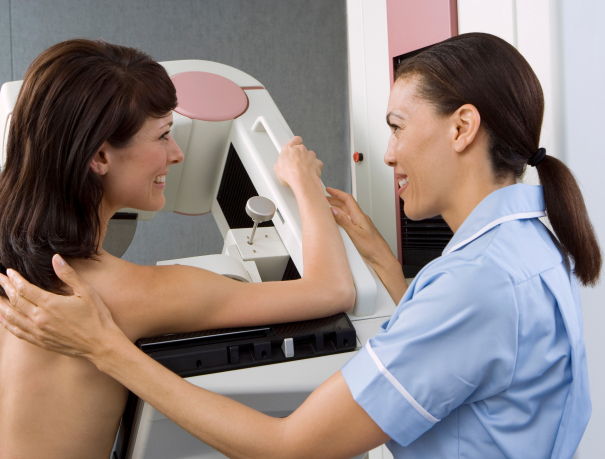
Mammography is the most used and most sensitive technique for the early detection of breast cancer, both for asymptomatic women and for those who have lumps, nipple discharge or sinking of the nipple. Using minimal radiation, mammography shows pictures of the inside of a woman’s breasts. It is so effective that it can show changes in the breast long before the doctor or patient notices it.
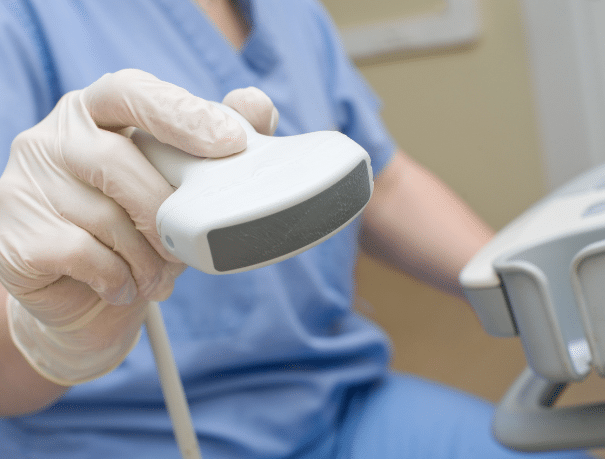
Breast ultrasound examines the breasts using ultrasound. It is a non-invasive test free of risks to women’s health, since there is no exposure to radiation. It allows to obtain images in real time to check the structure of the breast and characterize the findings obtained in the clinical examination or in mammography. Ultrasound alone is not indicated as a test for cancer detection, but is a complementary test.
Mammography, unlike ultrasound, can detect microcalcifications, small deposits of calcium that can warn of the presence of signs of breast cancer.
CASES IN WHICH IT IS RECOMMENDED
Who is it for?
Mammography is the most reliable and accurate method for early detection of breast cancer in asymptomatic women. It has been shown to reduce cancer mortality due to the finding of small tumors, when they are not yet palpable, and the chances of cure are greater. It is able to demonstrate the presence of clustered microcalcifications, which represent the most frequent form of presentation of breast cancer in its earliest stage. It is indicated as screening from the age of 35 in patients with no family history of breast cancer.
Ultrasound can be performed on women before 30 years of age, in pregnant or lactating women, and as a complement to mammography in the study of dense breast. It may be useful in patients who have silicone secretions or implants. Also in the follow-up of lesions that are only visible by ultrasound (not by mammography).
Instructions
Test preparation
Bilateral mammography is a non-invasive test that although it does not cause pain, can be a bit annoying, since to obtain the images the mammogram must compress the breasts. For the rest, it does not require preparation, it is fast and ambulatory, so at the end of the test you can resume your daily activities. During bilateral mammography, you should stand in front of the x-ray machine bare-chested and place a breast on a plate. Then, a second plate will press the breast against the first, crushing it firmly but without causing damage. The procedure is repeated with the other breast.
To perform a breast ultrasound, our medical staff will accommodate you on a stretcher and then perform a breast examination. It is a completely painless and minimally invasive test, so you just have to be calm and relax.

Medical professionals
The specialists who will assist you at CreuBlanca
A team of professionals to take care of you.

Related articles
CreuBlanca's blog
You will find from the hand of our professionals tips to improve your health and information on the latest technologies applied in the medical health sector.
 CreuBlanca
A Day with Dr. Garnica in the Operating Room: Inside Plastic and Reconstructive Surgery at CreuBlanca
 CreuBlanca
First Visit in Reproductive Medicine: How to Start the Process and What to Expect
 Health tips


