
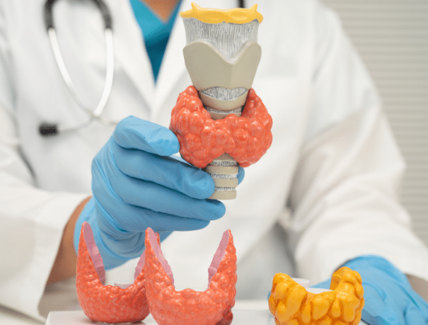
Thyroid scintigraphy
What does it consist of?
Scintigraphy is a nuclear medicine technique that involves the injection of small doses of radioactive material (tracer) to assess the function of different organs and systems.
The tracer, which emits gamma rays, travels through the bloodstream and is absorbed by the organs. Subsequently, a gamma camera with radiation detectors captures images of the highlighted areas.
In the case of thyroid scintigraphy, the tracer concentrates in the thyroid gland, allowing the evaluation of its size, shape, position and function. This test is particularly useful for studying thyroid activity, detecting the presence of nodules and determining whether they are hyperfunctioning or hypofunctioning.
When is it recommended?
Who is thyroid scintigraphy intended for?
Your doctor may recommend a thyroid scintigraphy in the following cases:
- To determine the cause of thyroid function disorders, such as hyperthyroidism or hypothyroidism.
- To assess the presence of thyroid nodules and analyse their activity (“cold” or “hot” nodules).
- To study the size, shape and location of the thyroid gland.
- To detect ectopic thyroid tissue or evaluate residual thyroid tissue after surgery.
- To monitor certain thyroid diseases or treatments.
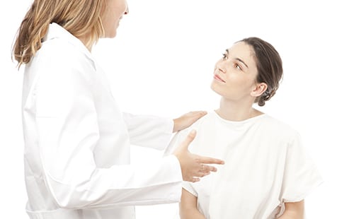
Preparation for the test
Instructions
Scans usually take about 3 hours. When you arrive, they will administer a contrast and until it takes effect, you cannot perform the test, which lasts between 10 and 15 minutes. While waiting for the reabsorption of the contrast, the patient can wait in one of our rooms or go for a walk around the center.

CREUBLANCA CENTERS
Find your nearest CreuBlanca center
Diagnosis Médica
Related articles
Expert opinion
You will find from the hand of our professionals tips to improve your health and information on the latest technologies applied in the medical health sector.
 Specialties
Cystitis and thrush in summer: how to tell them apart
 Specialties
Elevated PSA: the imaging that changes everything
 Specialties