
PET-CT
What does it consist of?
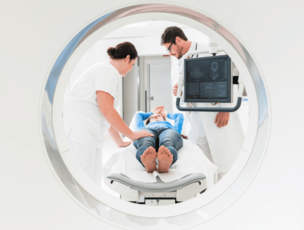
First the CT scan is performed and then the PET. CT scans allow images of slices or sections of the entire body using X-radiation. While PET allows to evaluate the metabolic activity of the cells present in the organs and tissues of the body by administering small doses of radioactive radiotracers. Subsequently, a computer joins the images so that they can be seen in three dimensions and thus have as much information as possible about what you want to study.
Before performing the test, we will perform a glycemic (blood sugar) control and then proceed to the administration of intravenous contrast. From this moment, you must remain approximately one hour at rest so that the contrast is distributed throughout the body evenly. Later, a radiologist technician will place you on the PET-CT table for the test. You should remain stretched on your back with your head resting on a cushion. Next, the table will be moved until the area to be studied is in the center of the PET-CT equipment.
CASES IN WHICH IT IS RECOMMENDED
Who is it for?
At CreuBlanca we carry out different types of PET-CT, among which are:
- Oncological. Early diagnosis, staging and monitoring of oncological processes, mainly in tumors of different types (urological, cardiological, neurological and pulmonary, among others).
- Cardiological. Visualize two defects attributable to a myocardial infarction, evaluate valvular and perivalvular involvement in prosthetic valve endocarditis, assess infectious involvement in intracardiac devices (pacemakers, ICDs, etc.), study large-vessel vasculitis and determine Sarcoidotic involvement in the cardiac muscle.
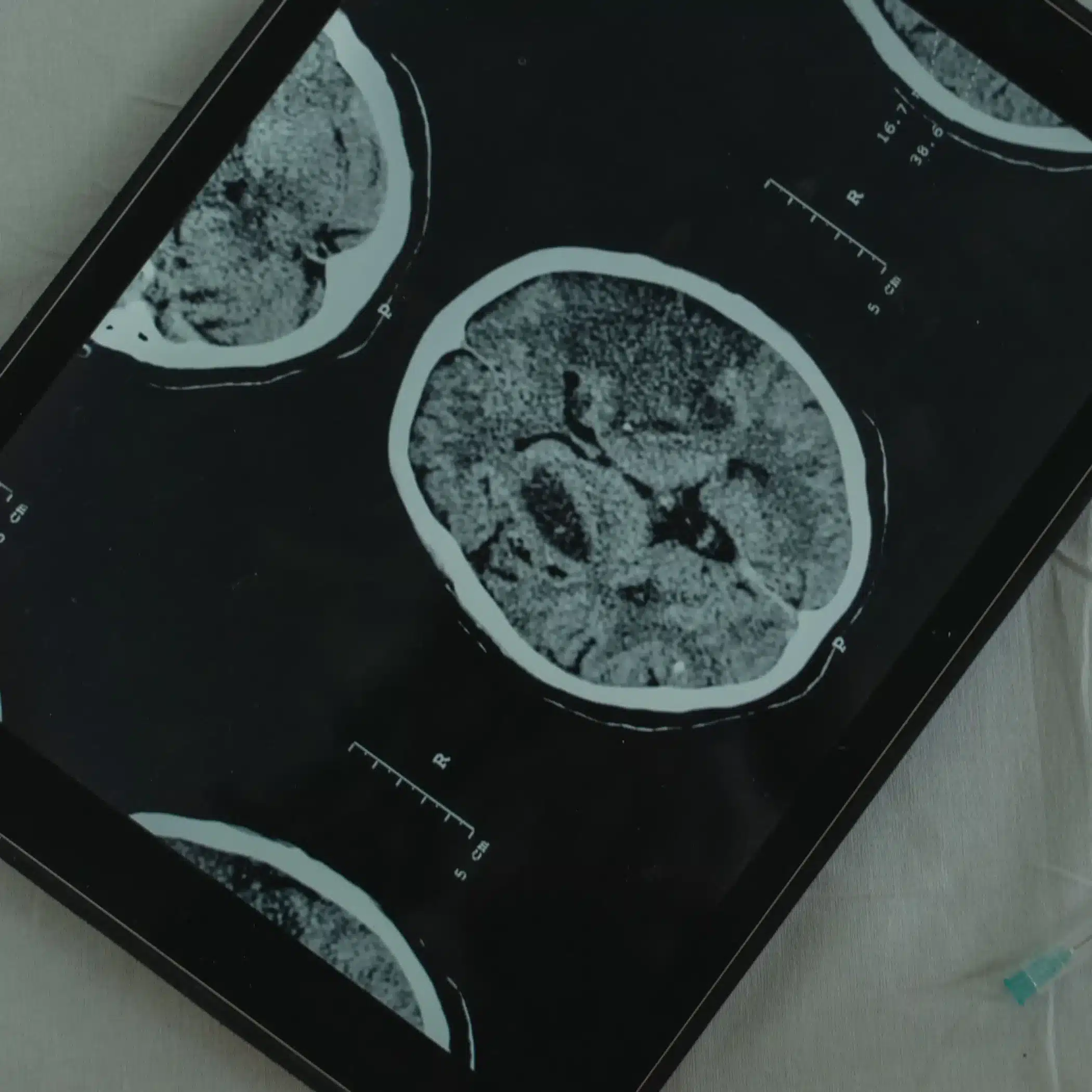
- Neurological. Early and differential diagnosis of different neurodegenerative diseases (dementia and atypical parkinsonism, Alzheimer’s, etc.).
INSTRUCTIONS
How should you prepare?
- Pregnancy: This test is contraindicated during pregnancy. For 24 hours after the test, you should stay away from pregnant women and children, as the radiopharmaceutical administered is radioactive.
- Fasting: You must go to the test with a minimum of 6 hours of fasting and drink approximately one liter of water. Although fasting is required, patients with chronic pathologies (kidney failure, hypertension, diabetes, etc.) can take their usual medication.
- Contrast administration: Before performing the test we will perform a glycemia (blood sugar) control and, then, we will proceed to the administration of intravenous contrast. In general, an analysis performed in the last 3 months is necessary to check that they have correct creatinine levels before the administration of iodinated contrast medium in patients with the possibility of renal failure.
- Breastfeeding: If you are breastfeeding, you should interrupt it for the next 24 hours. You should discard breast milk.
- Hydration: Drink plenty of water to remove contrast or radiopharmaceutical as soon as possible.

Medical professionals
The specialists who will assist you at CreuBlanca
A team of professionals to take care of you.


CREUBLANCA CENTERS
Find your nearest CreuBlanca center

Clinic CreuBlanca
Related articles
Expert opinion
You will find from the hand of our professionals tips to improve your health and information on the latest technologies applied in the medical health sector.
 Diagnostic Tests
What is the difference between an MRI, a CT scan, and a PET-CT scan?
 Specialties
Cystitis and thrush in summer: how to tell them apart
 Specialties