
Ultrasound pregnancy
What does it consist of?
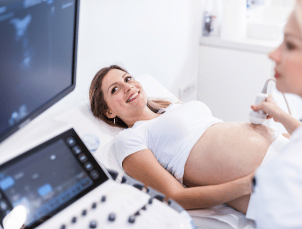
During the examination, the specialist moves the transducer over the abdomen, having previously applied the gel. This transducer sends sound waves (ultrasound) that “bounce” off different organs and tissues, forming the image.
- Ultrasound 12-14 weeks (first trimester): it is essential to detect the risk of chromosomopathies and review the general condition of the fetus, it also provides information on the characteristics of its vital organs. You will find out if it is a single or multiple gestation pregnancy, you will be able to listen to the rhythm of the heartbeat and see the size of the fetus. During the ultrasound, the risk of pathologies of chromosomal origin, such as Down syndrome, among others, is studied through certain parameters.
- Ultrasound 16-20 weeks (second trimester): it is essential to know the sex of the baby and rule out malformations. In addition, it evaluates the presence of alterations in the umbilical cord, amniotic fluid and placenta. During this ultrasound, if you wish, you will find out the sex of the future baby.
- Ultrasound 26-30 weeks (third trimester): The state and degree of maturation of the placenta, the amount of amniotic fluid and the state of the umbilical cord are studied. Also, the baby’s weight, size and position of the baby inside the uterus are estimated.
Cases in which it is recommended
To whom?
The pregnancy ultrasound is aimed at all those women who are in the gestation period, from 12 weeks and until late in the third trimester.

Instructions
How should you prepare?
This test does not require any specific preparation. You can lead a normal life.

Medical professionals
The specialists who will assist you at CreuBlanca
A team of professionals to take care of you.

CreuBlanca Centres
Find your nearest CreuBlanca center

Barcelona
CreuBlanca Tarradellas
Related articles
CreuBlanca's blog
You will find from the hand of our professionals advice to improve your health and information on the latest technologies applied in the medical health sector.
 Diagnostic Tests
24 Jul 2026
3 Min
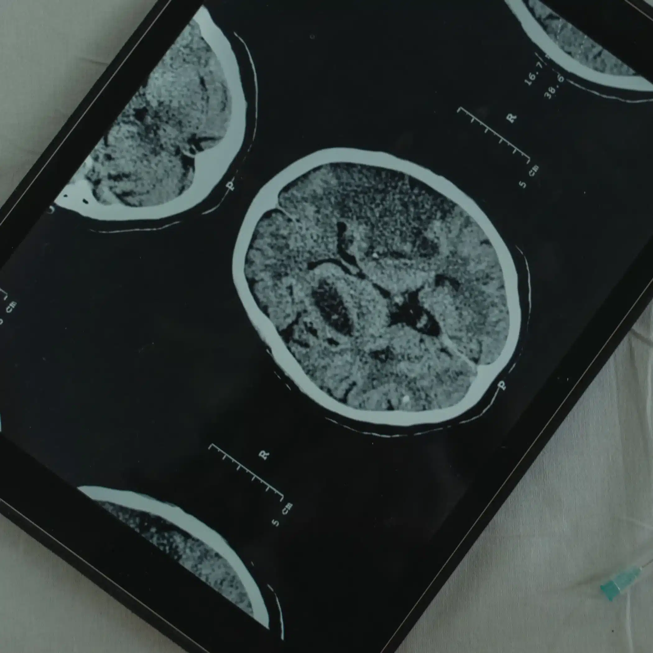
What is the difference between an MRI, a CT scan, and a PET-CT scan?
When it comes to making an accurate diagnosis, medical technology offers us various tools. It is very common to confuse tests such as MRIs, CT scans, and PET-CT scans, since they all allow us to see inside the human body. However, they use entirely different technologies and are prescribed for different purposes.
 Specialties
02 Jul 2026
3 Min
Cystitis and thrush in summer: how to tell them apart
Heat, humidity and long hours in a wet swimsuit make two very common conditions more frequent in summer: cystitis and vaginal thrush (candidiasis). Both affect the intimate area, both get worse with heat, and that is why many women confuse them.
 Specialties
22 Jun 2026
3 Min
Elevated PSA: the imaging that changes everything
Elevated PSA.
No pain, no symptoms, and no obvious signs that something might be wrong.