
MRI Cartilage Mapping / T1 and T2 Mapping
What does it consist of?
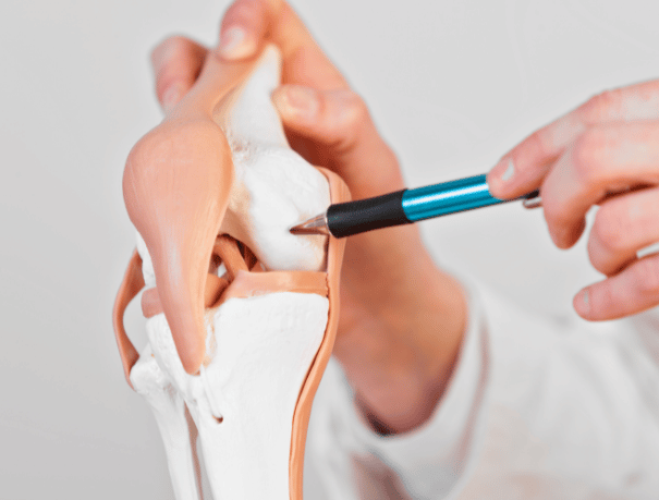
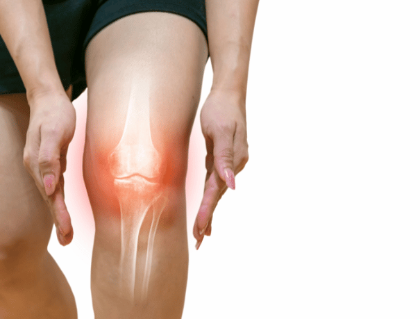
To carry out the test, you will have to lie face up with your head resting on a cushion and a radiologist will administer contrast to mark the area to be studied, generally the hip, knee, ankle or wrist. Next, the stretcher will move until the area of the body to be studied is in the center of the resonance equipment.
The test can last from 20 to 45 minutes. During this time, you will have to remain as calm as possible, since the taking of images is not continuous, but different image planes are made. Once finished you can continue with your daily life as normal.
Cases in which it is recommended
To whom?
The performance of MRI Cartilage Mapping is mainly indicated in the following cases:
- People who present degenerative cartilage pathologies, such as arthritis, allowing the application of an appropriate treatment for each case.
- Patients with chondral lesions of traumatic origin, that is, due to a fall or blow, and in which the cartilage has suffered small fissures, breaks and chondral detachments.
- People who have previously undergone surgical cartilage repair.
Instructions
How should you prepare?
- Contrast administration: This exploration requires intravenous contrast administration.
- Metal objects: The technician will give you the necessary instructions, provide you with a gown and ask you to remove any metal objects (jewelry, watch, piercings, hairpins, mobiles, dental prostheses and hearing aids).
- Heart devices: It’s important to tell the technician about any devices you’re implanted in your body (pacemakers, electrodes, and clips from previous surgeries).
- Hydration and lactation: Drink plenty of water to eliminate contrast from the body. If you are breastfeeding, you should stop for 48 hours. You should discard breast milk.

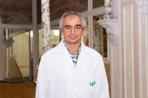
Medical professionals
The specialists who will assist you at CreuBlanca
A team of professionals to take care of you.

CreuBlanca Centers
Find your nearest CreuBlanca center

Clinic CreuBlanca

CreuBlanca Maresme Hospital
Related articles
CreuBlanca's blog
You will find from the hand of our professionals advice to improve your health and information on the latest technologies applied in the medical health sector.
 Specialties
Cystitis and thrush in summer: how to tell them apart
 Specialties
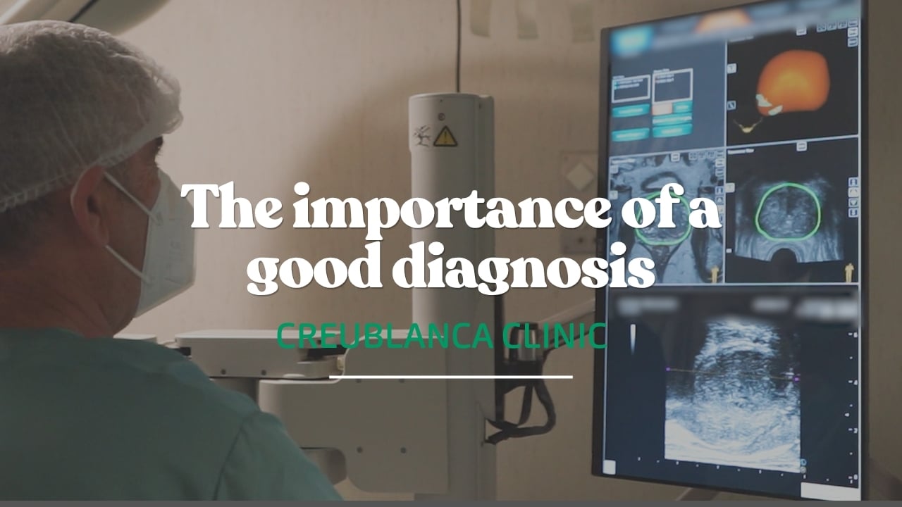
Elevated PSA: the imaging that changes everything
 Specialties